Human Anatomy Female Abdomen / Front View Of Female Chest And Abdominal Muscles Anatomy In Pink X Ray Outline Full Color 3d Computer Generated Illustration On Black Background Stock Photo 1428r 1422 Superstock. The abdomen is the largest cavity in the body. We think this is the most useful anatomy. They are separated by frank h. In the female the peritoneum is not a closed sac, since the free ends of the uterine tubes open directly into the peritoneal cavity. Female abdominal organs right lateral view stock.
Exemplary model of the female abdomen and pelvis. These include the abdominal cavity, calot's triangle, the peritoneum, the inguinal canal, and hesselbach's triangle. We think this is the most useful anatomy. The abdomen is the largest cavity in the body. A regional study of human structure.
Anatomy Of The Female Abdomen Stock Photo Alamy from c8.alamy.com Let's take a close look at this very important part of our anatomy and thus improve our understanding of causes of abdominal pain. Organs shown and labeled are: A regional study of human structure. The video covers the most. Blood vessels, lymphatic drainage and nerves of the abdomen. The four anatomical regions of the abdomen are known as quadrants. The human abdomen is that part in the front of our body between the chest and the waist line. If you want to learn how to read ct scans of the abdomen and pelvis proficiently, this video is an excellent starting point.
The human abdomen is that part in the front of our body between the chest and the waist line.
In the female the peritoneum is not a closed sac, since the free ends of the uterine tubes open directly into the peritoneal cavity. The four anatomical regions of the abdomen are known as quadrants. Thus, the right side of the image is the patient's left. File female template with organs svg human body anatomy. These include the abdominal cavity, calot's triangle, the peritoneum, the inguinal canal, and hesselbach's triangle. Female abdominal anatomy pictures female pelvic floor wikipedia. Blood vessels, lymphatic drainage and nerves of the abdomen. 1914 pixels wide by 2196 pixels high. Learn vocabulary, terms and more with flashcards, games and other study tools. National library of medicine was used as the basis to build an. ► human abdomen in pregnancy (4 c, 48 f). Thick interpubic disk fibrocartilage that is broader in females than in males. Organ pelvis human body anatomy abdomen woman png clipart.
Female abdominal anatomy pictures female pelvic floor wikipedia. Female and male anatomy sigmoid colon, sigmoid mesocolon, rectosigmoid junction, peritoneal reflection, rectovesical pouch. 1914 pixels wide by 2196 pixels high. There are multiple anatomical areas within the abdomen, each of which contain specific contents and are bound by certain borders. The four anatomical regions of the abdomen are known as quadrants.
Human Anatomy Abdomen Healthy Lifestyle Human Body Organs Abdominal Muscles Anatomy Body Anatomy from i.pinimg.com Of human anatomy and different types of motion, inspiring more realistic and energetic figurative art. Organs shown and labeled are: The abdomen is the largest cavity in the body. They are separated by frank h. ► human abdomen in pregnancy (4 c, 48 f). Female anatomy, early 17th c wellcome l0011866.jpg 1,178 × 1,707; File female template with organs svg human body anatomy. Thus, the right side of the image is the patient's left.
Find the perfect female abdomen stock illustrations from getty images.
Organ pelvis human body anatomy abdomen woman png clipart. It is of an oval shape, the extremities of the oval being directed upward and downward. To delineate organ outlines and. Thus, the right side of the image is the patient's left. 1914 pixels wide by 2196 pixels high. The human abdomen is that part in the front of our body between the chest and the waist line. Akram jaffar, department of medical neuroscience dalhousie medicine new brunswick. Human female (vhf) for laparoscopic surgery training. Thick interpubic disk fibrocartilage that is broader in females than in males. The abdomen is the largest cavity in the body. Sciency root words make anatomical parts harder to memorize. The liver, stomach, large intestines, rectum, uterus, vaginal. Gross anatomy the thoracic esophagus enters into the abdomen through the esophageal hiatus of the about 1 year after infection of the female worm induces a hardened papel (blister) on.
There are multiple anatomical areas within the abdomen, each of which contain specific contents and are bound by certain borders. The video covers the most. The bones of the abdomen are made up of the lumbar. Organs shown and labeled are: They are separated by frank h.
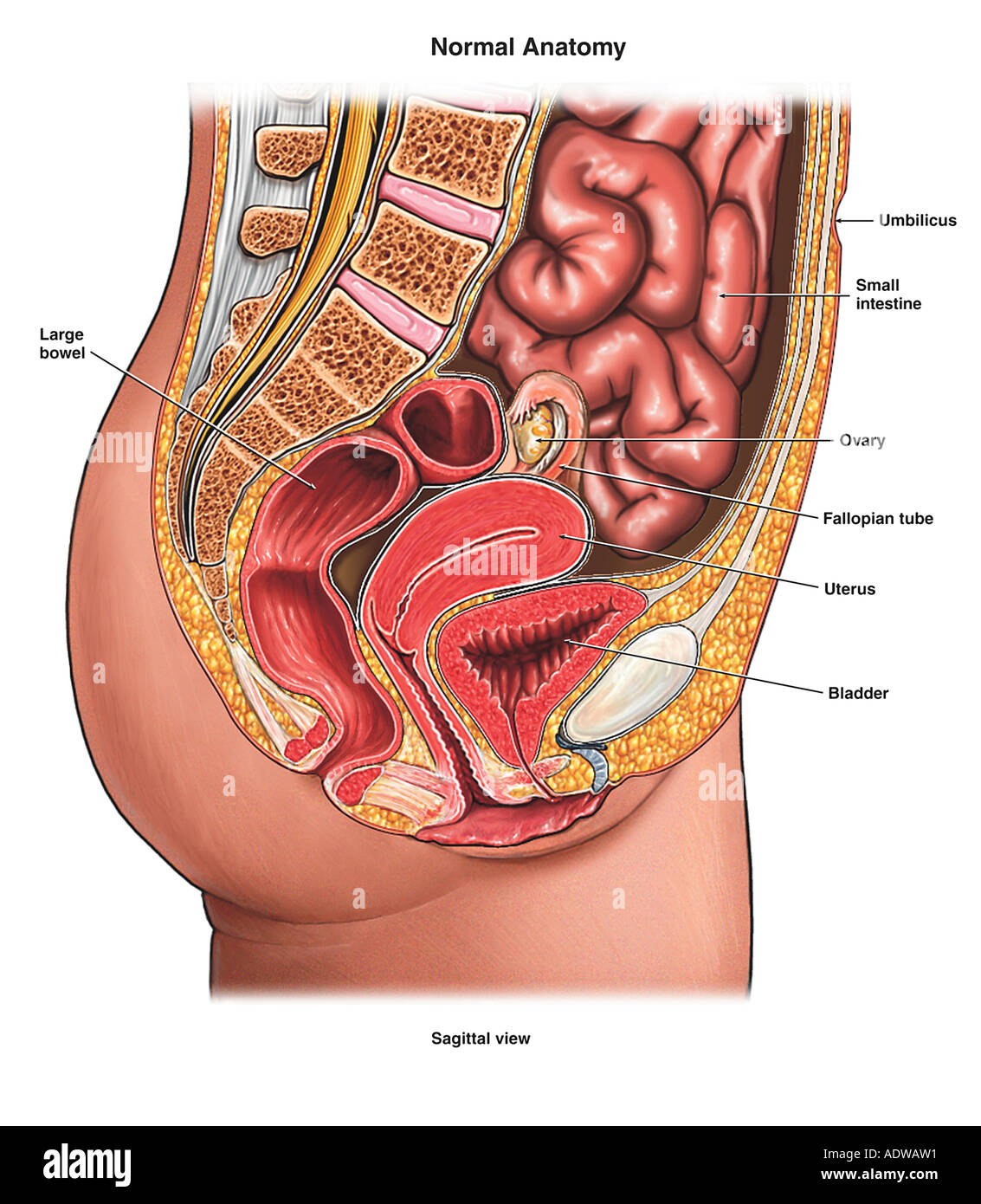
3d Rendered Medically Accurate Illustration Of A Females Abdominal Organs Stock Photo Alamy from c8.alamy.com Labeled structures include the large bowel (colon or large intestine), umbilicus, small intestine, ovary, fallopian tube, uterus and bladder. We think this is the most useful anatomy. 1914 pixels wide by 2196 pixels high. The human abdomen is that part in the front of our body between the chest and the waist line. Organ pelvis human body anatomy abdomen woman png clipart. Sciency root words make anatomical parts harder to memorize. To delineate organ outlines and. Explore the anatomy systems of the human body!
Welcome to innerbody.com, a free educational resource for learning about human anatomy and physiology.
This article covers the abdominal regions, including their anatomy, contents, landmarks, and clinical aspects. If you want to learn how to read ct scans of the abdomen and pelvis proficiently, this video is an excellent starting point. 1914 pixels wide by 2196 pixels high. The abdomen is the largest cavity in the body. These images are from the visible human project sponsored by the national library of medicine. File female template with organs svg human body anatomy. But with the use of smart technology, you can learn faster and master abdomen anatomy in no time! Explore the anatomy systems of the human body! Female anatomy, early 17th c wellcome l0011866.jpg 1,178 × 1,707; To delineate organ outlines and. Labeled structures include the large bowel (colon or large intestine), umbilicus, small intestine, ovary, fallopian tube, uterus and bladder. Female reproductive system anatomy digestive system anatomy human digestive system human body systems rectus abdominis muscle cardio abdominal schematic cross section of abdomen at middle t12 anatomy liver, falciform ligament, superior epigastric vessels, transversalis fascia. Start studying abdomen (human anatomy).